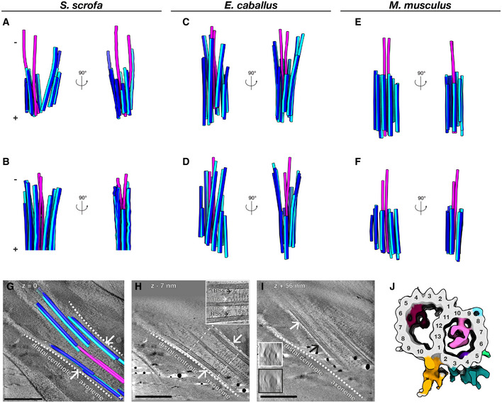
Figure 2. The distal centriole (DC) in mammalian sperm is composed of doublet microtubules arrayed asymmetrically around a pair of singlet microtubules.

-
A–FMicrotubules in the DC of pig (A, B), horse (C, D), and mouse (E, F) sperm traced from Volta phase plate cryo‐tomograms of intact sperm. Doublets are colored blue (A‐tubule in light blue, B‐tubule in dark blue), while singlets are pink.
-
G–ITomographic slices through cryo‐FIB‐milled lamellae of the DC‐to‐axoneme transition in pig sperm show how the change in geometry (G, white arrows and white dashed lines) coincides with the appearance of axoneme accessory structures (H, white arrows) and with density in the A‐tubule (I, compare insets in white and black boxes). In (I), the white and black arrows indicate where the cross‐sections in white and black boxes were taken from.
-
JIn situ structure of the pig sperm DC microtubule doublet with the tubulin backbone in gray and microtubule inner protein densities colored individually. The gold and turquoise densities on the luminal side of the doublet are consistent with the positions of parts of the inner scaffold. This structure represents the DC doublets closest to the axoneme (the area shown in (G–I)).
Data information: Labels: RSs—radial spokes, cpa—central pair apparatus, At—A‐tubule, Bt—B‐tubule. Scale bars: 250 nm.
