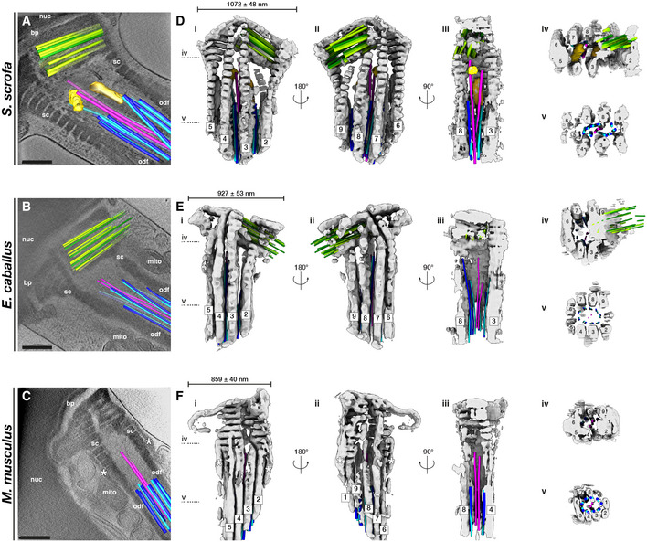
Figure 3. The connecting piece forms a large, asymmetric chamber around the sperm centrioles.

-
A–CSlices through Volta phase plate cryo‐tomograms of the neck region in intact pig (A), horse (B), and mouse (C) sperm. Proximal centriole triplets are shown in green, distal centriole doublets in blue (A‐tubule in light blue, B‐tubule in dark blue) and singlets in pink, and electron‐dense bars in yellow. Note the electron‐dense structures flanking the connecting piece in the mouse (asterisks in C, F).
-
D–FThree‐dimensional architecture of the flagellar base, with the connecting piece in gray, the proximal centriole in green, distal centriole doublets in blue (A‐tubule in light blue, B‐tubule in dark blue) and singlets in pink, and electron‐dense bars in yellow. The connecting piece was segmented semi‐automatically with a neural network, while microtubules were traced manually.
Data information: Labels: nuc—nucleus, bp—baseplate, sc—striated columns, odf—outer dense fibers, mito—mitochondria. Scale bars: 250 nm.
