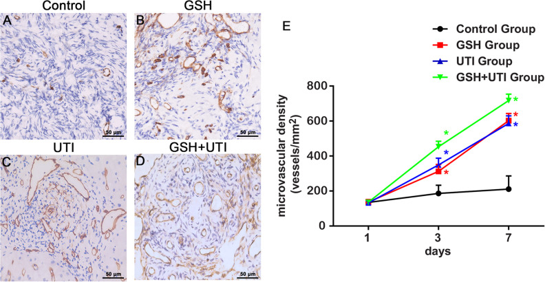
Figure 3.
Analysis of the murine microvessel in the human ovarian grafts. Immunohistochemical staining of murine microvessel in the human ovarian grafts from control group (A), GSH group (B), UTI group (C) and GSH+UTI group (D) at the 7th day after xenotransplantation. Curve graph (E) showed mMVD in human ovarian grafts from each group form at the 1st, 3rd and 7th day after xenotransplantation *Indication of statistical significance (P < 0.05) when compare to control group.

