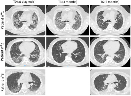
Figure 1.

Radiological findings of the three patients at the moment of the COVID‐19 diagnosis and after 3 (T3) and 6 months (T6) of follow‐up. The chest high‐resolution computed tomography (HRCT) showed an evolution of pneumonia. Patient 1, T0: presence of 40% ground glass pneumonia, T3: presence of 70% ground glass pneumonia and septal thickening, T6: presence of honey combing; Patient 2, T0: presence of 35% ground glass pneumonia, T3: improving of ground glass and parenchyma bands, T6: presence of 35% ground glass pneumonia; Patient 3, T0: presence of 25% ground glass pneumonia, T3: not available, T6: presence of 20% ground glass pneumonia and parenchymal bands
