Fig. 1.

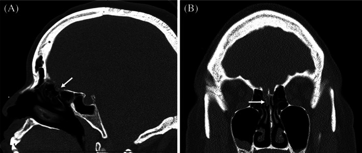
Computed tomography scan without contrast in the (A) sagittal plane and (B) coronal plane demonstrating the defect in the right cribriform plate (arrows).

Computed tomography scan without contrast in the (A) sagittal plane and (B) coronal plane demonstrating the defect in the right cribriform plate (arrows).