
FIGURE 4.
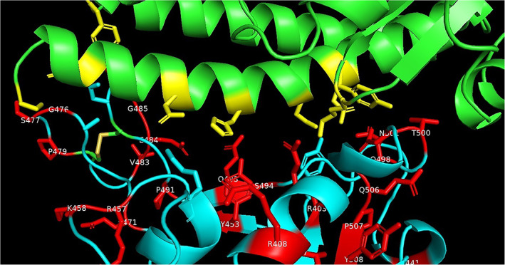
Interactions of spike protein residues (cyan) with ACE‐2 (green) side‐chain residues (yellow) that are within 3.2 Å in crystal structure of human SARS‐CoV‐2 spike protein RBD complexed with ACE‐2 receptor (PDB code: 6LZG). The spike protein mutated residues are shown in (red). PDB, Protein Data Bank; RBD, receptor binding domain [Color figure can be viewed at wileyonlinelibrary.com]