
Figure 1.
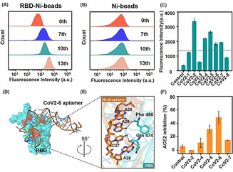
Aptamer CoV2‐6 identification and characterization. A,B) Flow cytometry to monitor the binding increment of enriched pools with RBD‐Ni‐beads (target beads) and Ni‐beads (control beads). C) Flow cytometry to investigate the binding performance of candidate sequences against SRBD. D) The results of molecular docking of overall structures of the CoV2‐6 aptamer (orange) and SRBD complex (SRBD is cyan and ACE2 binding amino acid residues are red), and E) the detailed analysis of the interface between CoV2‐6 aptamer and SRBD. F) The ACE2 inhibition efficiency of candidate sequences. SRBD was expressed by baculovirus‐insect cells.