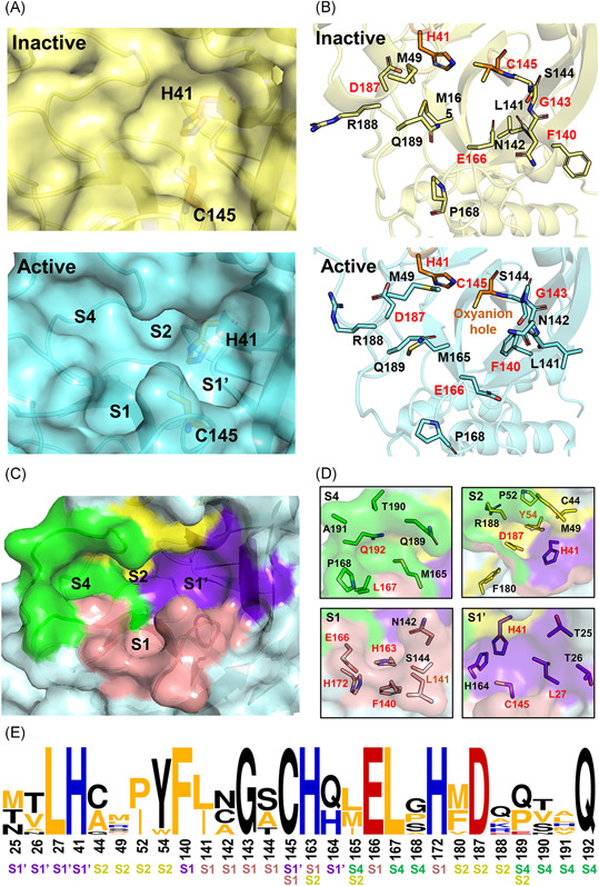
Figure 4.

Characterization of the substrate‐binding site of 3CLpro. (A) Expanded view of the substrate‐binding site of SARS‐CoV 3CLpro in its inactive and active forms (PDB ID: 1UJ1 and 2AMQ). (B) Detailed configurations of all residues involved in the binding site. Conserved residues are highlighted in red. (C,D) Residue configurations of each subsite of SARS‐CoV‐2 3CLpro (PDB ID: 6LU7). (E) The sequence logo69, 70 representation of the conservation of residues forming the binding site. The color coding is defined according to the chemical properties of amino acids (polar or neutral: black, basic: blue, acidic: red, and hydrophobic: orange) and the subsite legend is given under each amino acid [Color figure can be viewed at wileyonlinelibrary.com]
