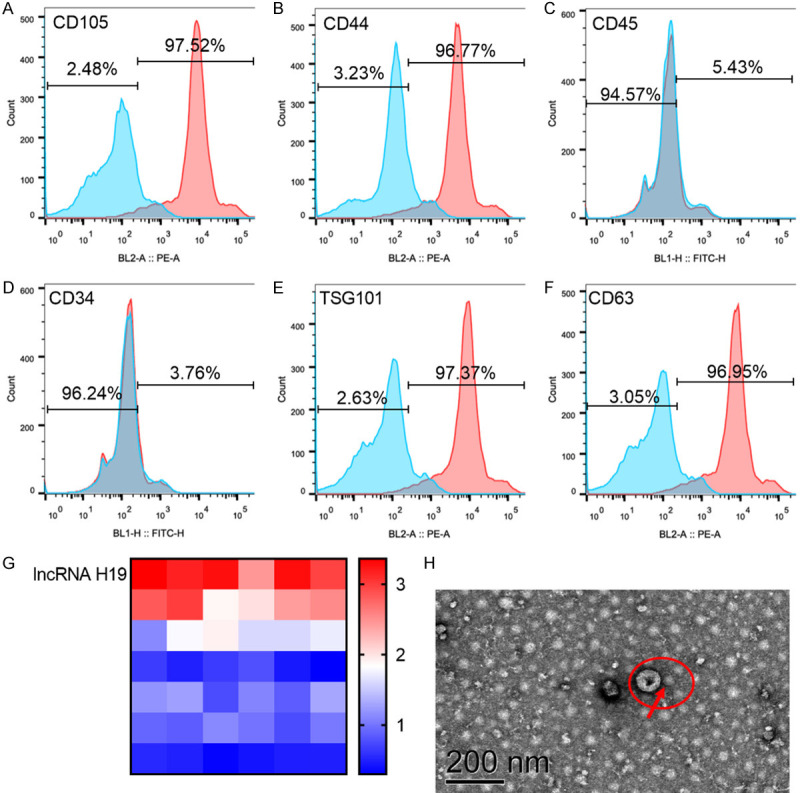
Figure 3.

Determination of mesenchymal stem cells and exosomes. A-D: Flow cytometry was used to analyze the surface markers CD105, CD44, CD45 and CD34 of mesenchymal stem cells, in which CD105 and CD44 were positive, while CD45 and CD34 were negative. E, F: Exosome surface markers TSG101 and CD63 isolated from mesenchymal stem cells were analyzed by flow cytometry, and TSC101 and CD63 were positive. G: LncRNA H19 was highly expressed in exosomes isolated from mesenchymal stem cells. H: Electron micrograph of exosome, with the scale of 200 nm.
