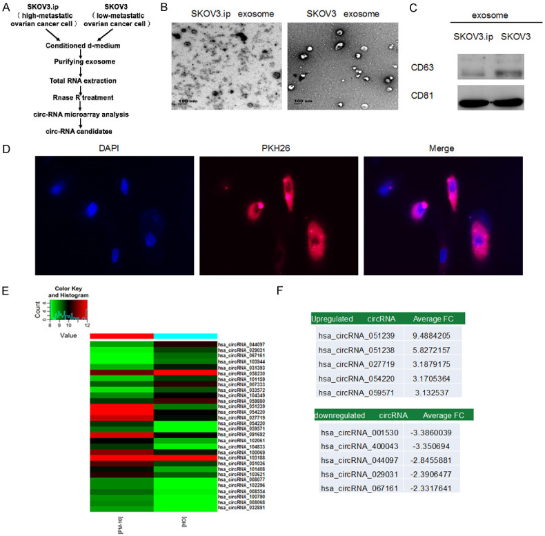
Figure 1.

The identification and features of circRNA in tumor exosomes. A. Schematic screening workflow of circRNA candidates. B. TEM image of exosomes isolated from SKOV3.ip (high-metastatic ovarian cancer cell) and SKOV3 (low-metastatic ovarian cancer cell) cell lines. C. Exosomes from culture media were examined by WB (CD63 and CD81). D. SKOV3 cells were treated with 5 μg of PKH26-labeled exosomes (red) derived from SKOV3.ip cells. The nuclei were visualized by DAPI staining. Red: PKH26; Blue: DAPI (nuclei). Confocal microscopy was used to image fluorescent cells. E. Heat map of differentially expressed circRNAs. ‘Red’ and ‘green’ indicates high and low relative expression, respectively. F. The top five upregulated and down-regulated circRNAs.
