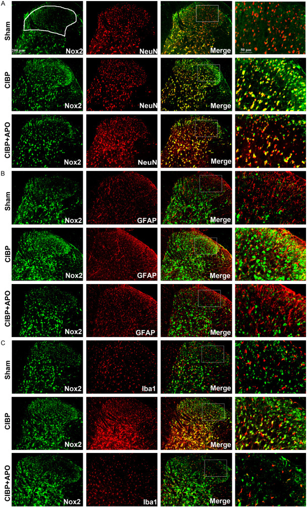
Figure 5.
Expression and cellular localization of Nox2 in the ipsilateral spinal cord. A-C. Nox2 (green) double fluorescence labeling with NeuN (red) for neurons, GFAP (red) for astrocytes and Iba1 (red) for microglia in the spinal cord dorsal horn at day 21 after tumor cell implantation. Amplified pictures showed the co-localization of Nox2 (green) and NeuN (red), GFAP (red) or Iba1 (red). B. The results showed that Nox2 was upregulated in CIBP + DMSO group compared with sham + DMSO group. C. Repeated injection of APO (200 mg/kg, i.p.) for 5 consecutive days notably decreased the expression of Nox2. The results showed that Nox2 was co-localized mostly with microglia (yellow) and neurons (yellow). (***P < 0.001 compared with the sham + DMSO group. ###P < 0.001 compared with the CIBP + DMSO group. n = 3 in each group). Scale bar = 200 μm.

