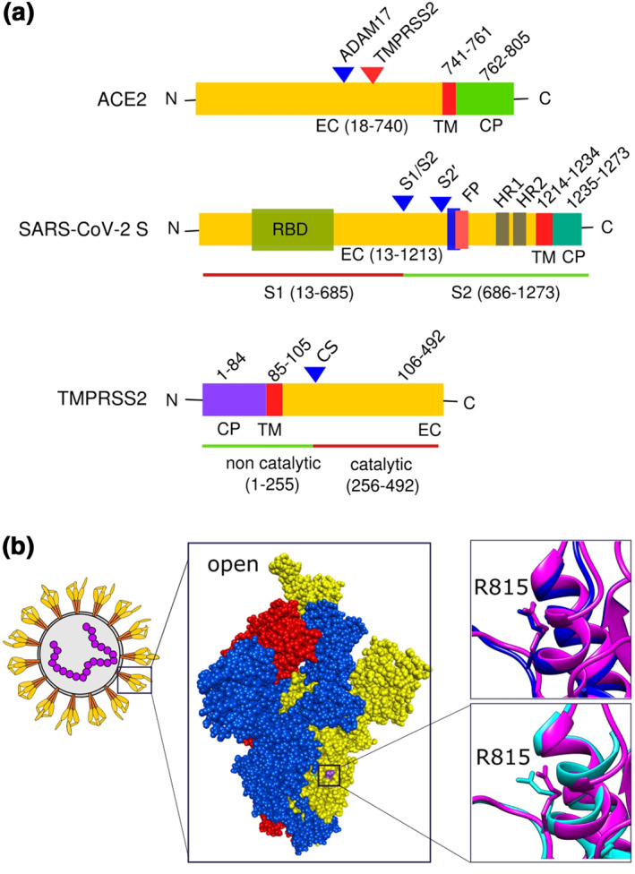
FIGURE 2.

Domains of important proteins and ACE2 binding mediated virus spike protein conformational shift: (a) domains of ACE2, SARS‐CoV‐2 spike protein and TMPRSS2; (b) spike protein trimer of SARS‐CoV‐2 (PDB ID 6XM3) with one monomer open (yellow), and two closed (blue and red). Two superimposed images shown to depict S2′ cleave site residue arginine 815. Closed (blue, PDB ID 6ZGE) and open (pink, PDB ID 6XM3) spike protein monomer superimposed both without ACE2 bound (right top). Open no‐ACE2‐bound spike protein (pink, PDB ID 6XM3) was superimposed on open with ACE2 bound (cyan, PDB 7A96) (right bottom). Discovery Studio Visualizer v17.2.0 and Chimrea v1.13.1 were used to visualize and superimpose S proteins, respectively. Inkscape v0.92.3 was used to compile the figure. CP, cytoplasmic; CS, autocatalytic site; EC, extracellular; FP, fusion peptide; HR, heptad repeat; TM, transmembrane
