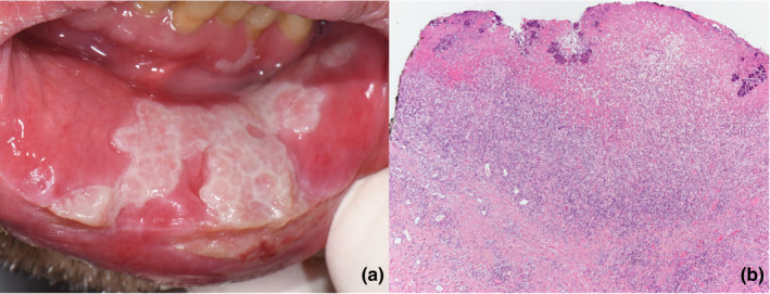
FIGURE 1.

(a) Painful ulcerated plaque of the mucosal side of the inferior lip. Similar lesions affected both margins of the tongue, both lips, and soft palate. (b) At low power, the oral mucosa was ulcerated with granulation tissue and fibrino‐leukocytic material including bacterial colonies. Dense inflammatory infiltrate was present in the submucosa
