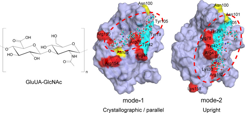
Figure 2.
HA molecule and two binding modes, mode-1 and mode-2, of HA on the CD44 HABD surface. Red, yellow, and blue colors on molecular surface denote positively charged (Arg and Lys), amidic (Asn), and aromatic (Tyr) residues, respectively, which were supposed to be important for the interaction with HA in each binding mode. The blue, white, red, and navy balls denote carbon, hydrogen, oxygen, and nitrogen atoms, respectively, in the HA molecule. The figures of the center and right were reprinted with permission from ref (11). Copyright 2004 Elsevier

