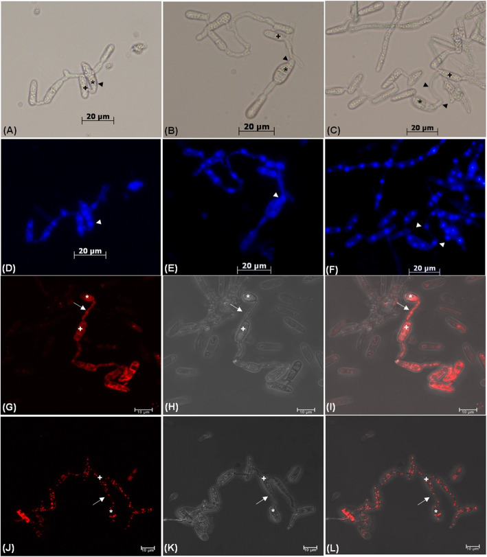
Fig. 6.
Transfer of nucleus and different cell organelles during CAT fusion between C. gloeosporioides and C. siamense. a, b and c Bright-field microscopy of CAT fusion between the 17 days old conidia C. gloeosporioides and C. siamense. d, e and f Visualization of nuclear transfer between the 17 days old conidia of C. gloeosporioides and C. siamense by DAPI staining using fluorescence microscope. g Fluorescent, h Brightfield and i Overlapping microscopic images of mitochondrial movement by MitoRed staining. j Fluorescent, k Brightfield and l Overlapping microscopic images of movement of lipid droplets by Nile red staining. Arrowhead indicates nucleus, arrow indicates CAT fusion, asterisk (*) symbol indicates C. siamense conidia and plus (+) symbol indicates conidia of C. gloeosporioides. Scale bar = 20 μm for images (a-f) and 10 μm for images (g-l)

