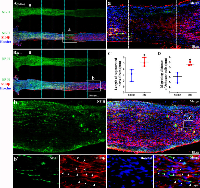
Fig. 6.
The in vivo effect of Btc recombinant protein after rat sciatic nerve transection and silicone bridging. Representative immunofluorescence images of rat sciatic nerve segments treated with (A) saline control or (B) Btc recombinant protein at 10 days after nerve transection and silicone bridging. Green color indicated NF-H, red color indicated S100β, and blue color indicated nucleus. Boxed areas in (A and B) were shown in higher magnifications in (a and b), respectively. (b’) was from boxed area of (b). Arrows indicated the regeneration site. Arrowheads indicated Schwann cell-formed cord in the nerve bridge. Scale bars indicated 1000 µm in (A and B) and indicated 100 µm in (a and b) and 20 µm in (b’). (C and D) Averaged (C) length of regeneration nerve fibers and (D) migrating distance of Schwann cells summarized from three experiments. Statistical evaluation was performed using t-test. *p-value < 0.05 versus saline control

