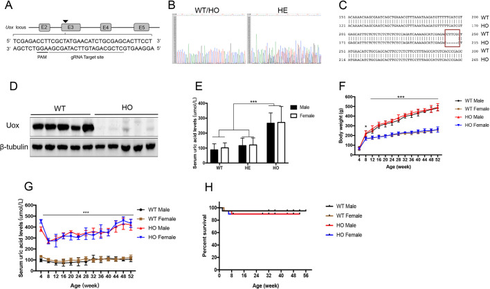
Fig. 1.
Generation of UOX-knockout (KO) rats with hyperuricemia. (A) Schematic representation of UOX KO using CRISPR/Cas9. (Top) Uox locus, boxes indicate exons and connecting lines indicate introns. (Bottom) Portion of the Uox sequence, showing the gene editing site (gray underlined), and the protospacer adjacent motif (PAM) region (black underlined). (B) Sanger sequencing of the PCR-amplified genomic Uox locus revealed DNA disruption at the gRNA-target site downstream of the Cas9 cleavage site in rats heterozygous (HE) for Uox KO, but not in wild-type (WT) rats or in rats homozygous (HO) for Uox KO. (C) Schematic targeting and deep sequencing reads of the Uox-locus with deletions (red box) in HO rats. (D) Hepatic expression of UOX protein determined by western blotting (8-week-old males, n=5). (E) Serum urate levels of WT, HE and HO rats, of both sexes (8 weeks of age, n=5 per group). (F) Body weight of male and female WT or HO rats (aged between 4 and 52 weeks), measured every 4 weeks (n=5 per group). (G) Serum urate levels of male and female WT and HO rats (aged between 4 and 52 weeks), measured every 4 weeks (n=5 per group). (H) Kaplan–Meier plot, showing the survival rates of HO (male, n=50; female, n=50) vs WT rats (male, n=50; female, n=50) from birth to 56 weeks of age. Values are presented as the mean±s.d. *P<0.05, ***P<0.001.

