Fig. 3.
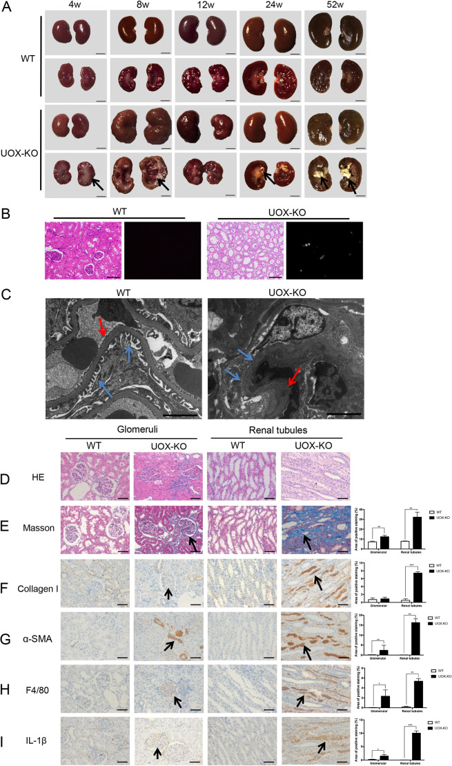
Renal histopathology was impaired in UOX-KO rats. (A) Images show complete kidneys and longitudinal sections of kidneys from male UOX-KO and WT rats as indicated, in weeks (w). Arrows either indicate lesions (4w and 8w) or urate crystals (24w and 52w) in kidneys of UOX-KO rats. (B) H&E staining (right) and polarized light images (left) of kidneys from 16-week-old WT and UOX-KO rats. Urate crystals, detected under polarized light in the kidney of the UOX-KO rat. (C) Transmission electron microscopy of kidney samples from male WT and UOX-KO rats at 8 weeks of age. Blue arrows indicate podocytes, red arrows indicate the basement membrane. (D–I, left) Images show kidney tissue collected from 12-week-old WT or UOX-KO rats. Columns one and two show glomeruli, columns three and four show renal tubules. Tissue was analyzed using H&E staining (D) or Masson's trichrome staining (E), or immunostained for the renal expression of collagen I (F), alpha-smooth muscle actin (α-SMA) (G), F4/80 (H) and interleukin (IL)-1β (I) by using corresponding antibodies (n=5 per group). Arrows in E–I indicate positive staining. Scale bars: 0.5 cm (A), 200 μm (B), 2 μm (C), 50 μm (D–I). (Right) Bar graphs showing quantification of each analysis. Values are presented as the mean±s.d. *P<0.05, **P<0.01, ***P<0.001.

