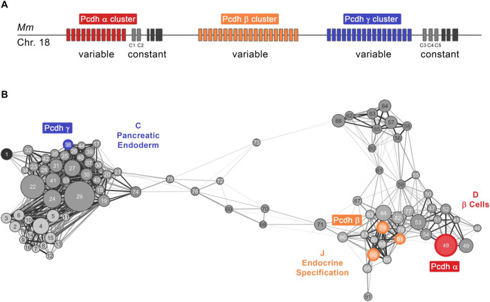
Fig. 3.
Distribution of clustered protocadherins in β-cell development GCN. (A) Schematic of genomic DNA locus (mouse chromosome 18) containing clustered protocadherins (Pcdh) genes organized in three closely linked gene clusters designated α, β and γ. Variable exons of the Pcdhα cluster sub-family are colored in red, Pcdhβ in orange and Pcdhγ in blue. Homologous Pcdhα and Pcdhγ C family variable exons are in light gray and constant exons are shown in black. (B) Different cluster sub-families of protocadherins are found in modules (square nodes) falling into meta-modules representing different developmental parts of the network. A meta-network view of GCN highlights that the module containing Pcdhα genes (red) is in meta-module D (mature β-cells), modules with Pcdhβ genes (orange) are in meta-module J (endocrine specification), and a module with Pcdhγ genes (blue) is in meta-module C (pancreatic endoderm).

