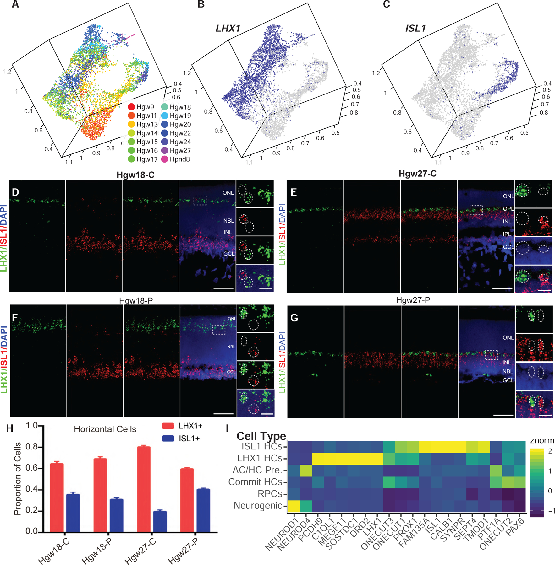
Figure 5. Identification and Differentiation of Two Horizontal Cell Subtypes within the Developing Human Retina.

(A–C) UMAP embedding of horizontal cells, colored by (A) age or (B–C) relative expression of (B) LHX1 and (C) ISL1.
(D–G) RNAScope detecting expression of LHX1 and ISL1 transcripts in central (D and E) and peripheral (F and G) Hgw18 and Hgw27 human retina, with higher-magnification views of the boxed regions. Nuclei are counterstained with DAPI. Scale bar, 50 μm and 10 μm (magnified views).
(H) Quantification of the proportions of each horizontal cell subtype in central and peripheral retina at ages Hgw18 and 27 from fluorescent in situ hybridization experiments. Data are mean ± SEM.
(I) Heatmap showing relative cell type expression of horizontal cell commitment, differentiation, and subtype specification genes. Abbreviations: Hgw, human gestational weeks; Hpnd, human postnatal day; C, central retina; P, peripheral retina; NBL, neuroblast layer; GCL, ganglion cell layer; ONL, outer nuclear layer; OPL, outer plexiform layer; INL, inner nuclear layer; IPL, inner plexiform layer; AC/HC Pre., amacrine cell/horizontal cell precursors; Commit HCs, committed horizontal cells; ISL1 HCs, ISL1-positive horizontal cells; LHX1 HCs, LHX1-positive horizontal cells.
