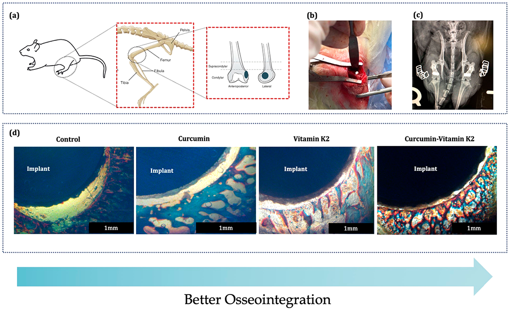
Figure 8.

In vivo surgical procedure and bone bonded zone shown by Masson Goldner staining: (a) 3/5 mm defect location at the lower epicondyle of the rat distal femur model, (b) implantation procedure, (c) postsurgical radiograph showing the placement of implant, and (d) Masson Goldner staining showing the implant and surrounding bone after sacrificing the rats at 5 days (n = 4).
