
Figure 9.
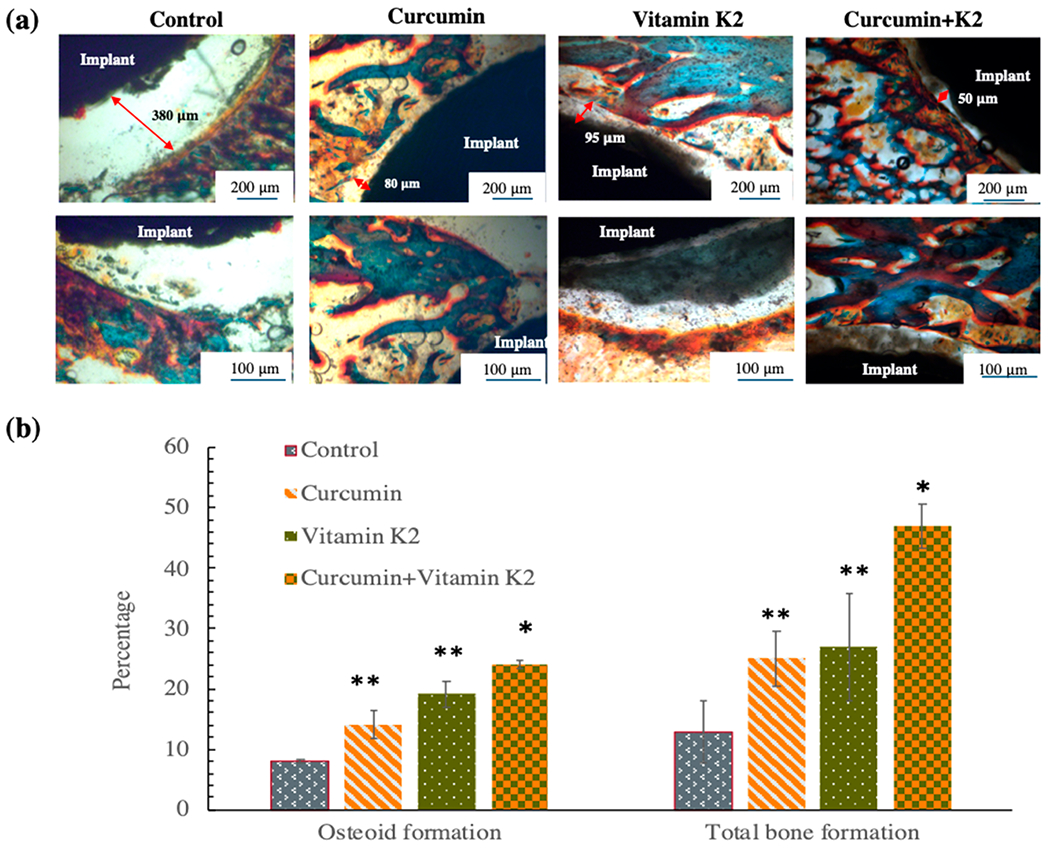
(a) In vivo Masson Goldner staining by curcumin-, vitamin K2-, and curcumin + vitamin K2-loaded HA-coated Ti implant showing the gap between the implant and the surrounding bone tissue after 5 days of implantation at rat distal femur model. The new bone formation or osteoid tissue is stained with red/orange; mineralized bone tissue is stained with green/blue, and the implant can be seen in black. (b) Histomorphometric analysis by ImageJ software showing percentage osteoid or new bone tissue formation and percentage total bone formation within 250 μm radius of implant after 5 days. Drug-loaded implant exhibited statistically significant difference in osteoid formation and total bone formation after 5 days indicating better osseointegration ability compared to the control (*p < 0.001, **p < 0.05).