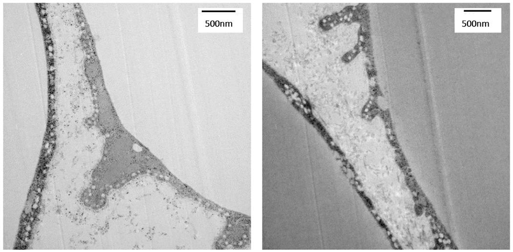
Figure 2: Electron microscopy of closly interacting adipocytes and macrophages: invaginated lipid structures budding off adipocytes and many vesicular structures resembling exosomes.

Adipose tissue was collected from lean chow fed C57BL/6J mice, cut into 1 mm pieces, and immediately fixed in 2.5% glutaraldehyde, before being prepared by Vanderbilt Imaging Core for TEM imaging. Images were captured on a Philips/FEI T-12 transmission electron microscope.
