
Figure 1.
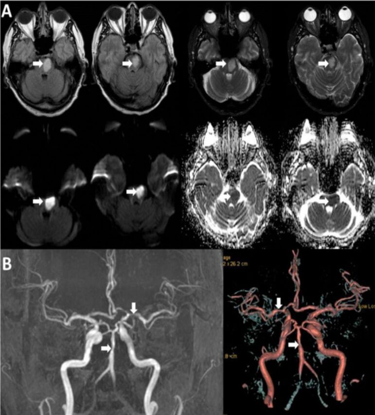
Neuroimaging studies of our patient with A) MRI of the brain showing acute infarction at the left aspect of the pons/lower midbrain with high signal intensity on FLAIR and T2-weighted images and restriction of diffusion with minimal mass effect and, B) MRA showing multiple segments with irregularity and narrowing at the basilar artery and left M-1 segment.