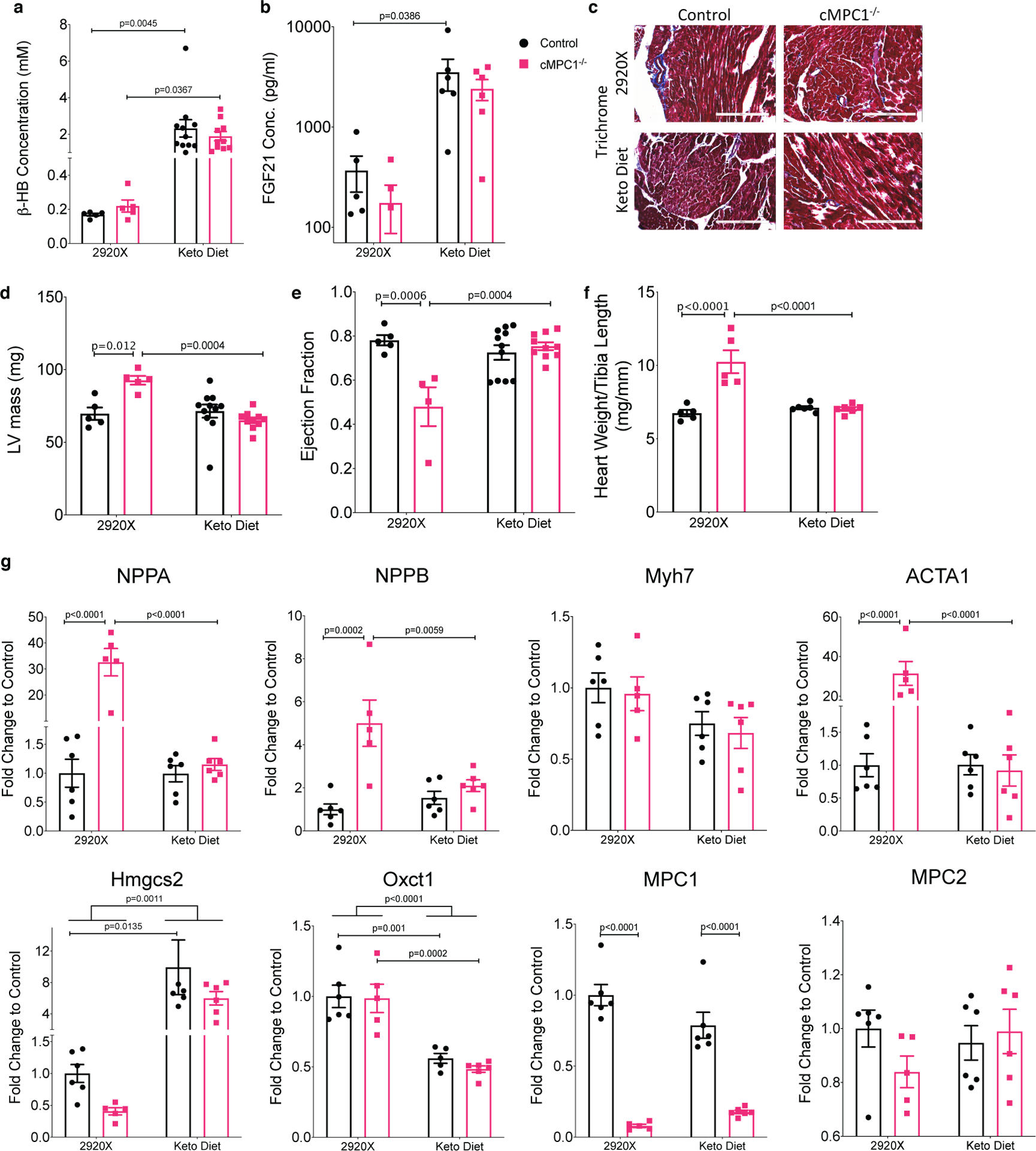
Figure 5. Ketogenic diet feeding prevents cardiac dysfunction in cMPC1−/− mice.

Control and cMPC1−/− mice were fed with normal chow (2920X) or a ketogenic diet at the age of 3 weeks. (a-b) serum concentration of β-HB (a) and FGF21 (b) after 8 weeks of feeding. (c) Trichrome staining reveals no increase in cardiac fibrosis in cMPC1−/− mouse hearts. Scale bar, 200 μm. Images are representative of n= 3 per group. (d-e) LV mass (d) and ejection fraction (e) were determined by echocardiography after 15 weeks of feeding. (f-g) Heart weight/Tibia length (f) and gene expression analysis (g) were determined at the time of sacrifice after 18 weeks of feeding. For panels a and d-f, n=5 (Control-2920X), n=5 (cMPC1−/−−2920X), n=11 (Control-Keto), n=10 (cMPC1−/−-Keto); for panel b, n=5 (Control-2920X), n=5 (cMPC1−/−−2920X), n=6 (Control-Keto), n=6 (cMPC1−/−-Keto); for panel g, n=6 (Control-2920X), n=5 (cMPC1−/−−2920X), n=6 (Control-Keto), n=6 (cMPC1−/−-Keto). Data are presented as mean ± SEM and P value was determined by two-way ANOVA followed by Tukey multiple comparison test.
