Figure 1.

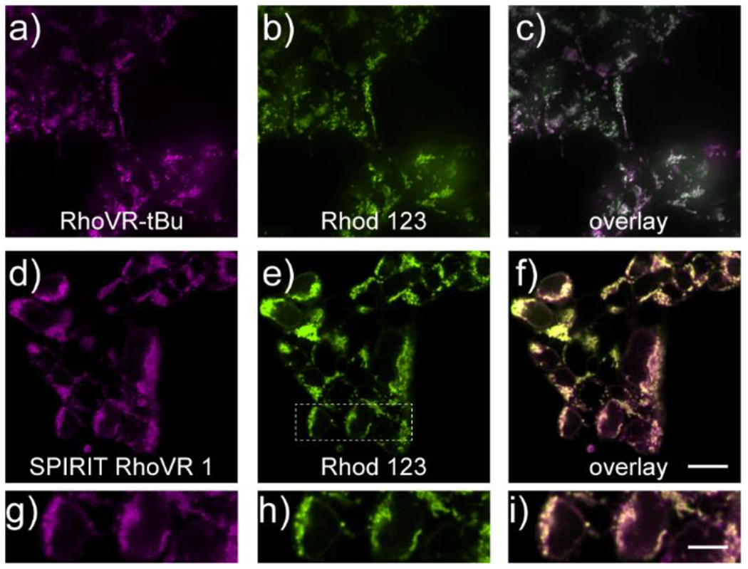
SPIRIT RhoVR 1 localizes to mitochondria in mammalian cells. Confocal fluorescence microscopy images of HEK cells stained with either a) RhoVR-tBu (250 nM) or d) SPIRIT RhoVR 1 (250 nM) and rhodamine 123 (250 nM, b and e). Overlay image of rhodamine 123 and either c) RhoVR-tBu or f) SPIRIT RhoVR 1. g-i) Expanded view of the boxed region in panel (e). Scale bar is 20 μm (a-f) or 10 μm (g-i)
