Figure 2.
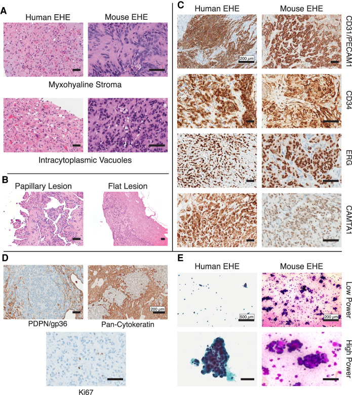
Mouse EHE is histologically identical to human EHE. (A) Representative H&E photomicrographs of both human and mouse EHE displaying the hallmark histologic features of EHE myxohyaline stroma and intracytoplasmic vacuoles. Scale bars, 50 µm. (B) Representative H&E photomicrograph of murine EHE displaying the two morphological phenotypes of peritoneal surface tumors. Scale bars, 50 µm. (C) Immunohistochemical staining of mouse and human EHE for defining IHC stains. The primary antibodies used are listed at the right. Scale bars, 50 µm unless specifically listed. (D) Immunohistochemical staining of mouse EHE for the defining IHC stains. The primary antibodies used are listed below. Scale bars, 50 µm unless specifically listed. (E) Cytology of human and murine EHE at low and high magnification (Left) Human EHE from a pleural effusion (Papanicolaou stain). (Right) Cytopathology of malignant ascites from mouse EHE (Diff Quik staining). Scale bars, 50 µm unless specifically listed.

