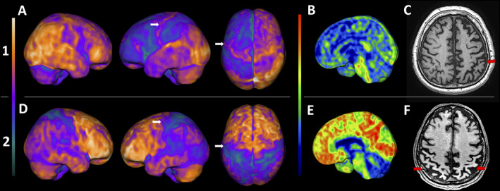
FIG. 1.

Upper row (1) patient 1: (A) FDG‐PET 3D‐stereotactic surface projection (3D‐SSP, software cortex ID suite, GE Healthcare): asymmetric frontoparietal hypometabolism, including the sensory and motor cortex, worst on the left side. PIB‐PET 3D‐SSP (B) negative for amyloid deposition, and T1‐weighted MRI (C) with asymmetric frontoparietal atrophy, also worst on the left (red arrows). Lower row (2) patient 2: (C) FDG‐PET 3D‐SSP: asymmetric posterior temporoparietal hypometabolism, also including the sensory and motor cortex, worst on the left side. PIB‐PET 3D‐SSP (E) positive for diffuse cortical amyloid deposition, and T1‐weighted MRI (F) showing bilateral parietal cortical atrophy (red arrows). The white arrows on A and D point to the supplementary motor cortex, probably related to the synkinesis.
