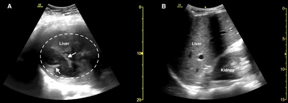
FIGURE 1.

Point‐of‐care ultrasound of the right upper quadrant in the coronal plane demonstrating (A) multiple hypoechoic collections and septations (white arrows) within the liver parenchyma consistent with a pyogenic liver abscess (dotted white circle) and (B) a representative example of normal liver parenchyma echotexture
