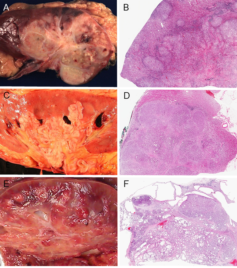
FIGURE 2.

Gross features and low power images. Gross features (A, C, E). (A) Renal medullary carcinoma (RMC): poorly circumscribed tumor occupying a large part of the parenchyma. (C) Collecting duct carcinoma (CDC): tumor centered in the medulla. (E) FH-deficient RCC: poorly-defined tumor involving the cortex and medulla. Low power images (B, D, F). (B) RMC: the tumor displays infiltrating irregular border and satellite nodules are seen in the cortex. (D) CDC: the tumor is overall well-defined, but focally invasive. Infiltrating growth pattern is also seen. (F) FH-deficient RCC: The tumor is overall well circumscribed with tubulocystic pattern. Multiple benign cystic lesions are seen in adjacent uninvolved renal parenchyma.
