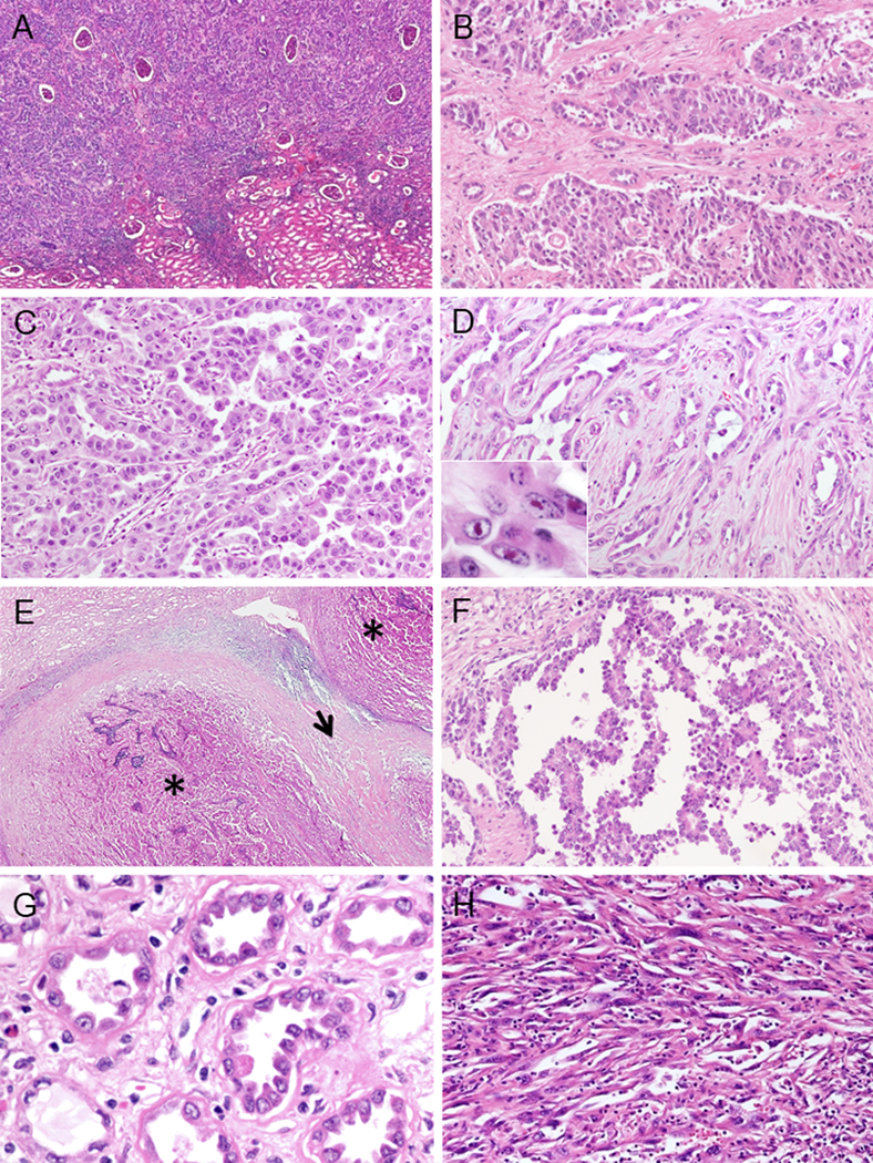
FIGURE 4.

Morphological patterns of CDC (A-F). (A) Interstitial infiltrating growth. (B) Solid sheets-nested pattern. (C) Tubulopapillary pattern. (D) Infiltrating glandular pattern. Small or medium sized elongated tubules in desmoplastic stroma. In7set shows prominent nucleoli. (E) Multinodular infiltrating growth pattern. Multiple infiltrative nodules of varying size with papillary architecture (*) and desmoplasia between nodules (arrow). (F) Intracystic papillary pattern with delicate fibrovascular core. Other findings of CDC (G-H). (G) Dysplastic in situ change within adjacent collecting ducts. (H) Sarcomatoid change.
