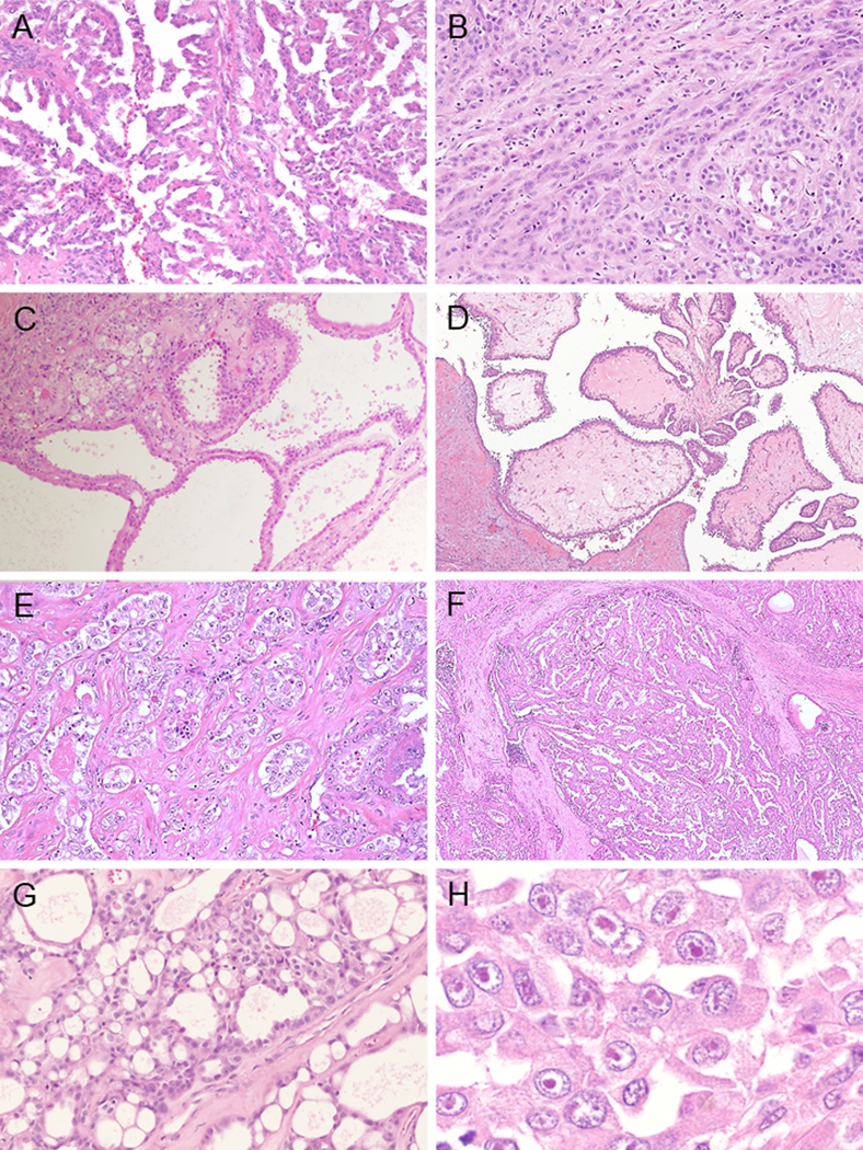
FIGURE 5.

Morphological pattern of FH-deficient RCC (A-H). (A) Papillary pattern. (B) Cord-like or small nests architectures. (C) Tubulocystic pattern. Poorly differentiated foci are identified in left upper sides. (D) Intracystic papillary pattern with hyalinized cores. (E) Infiltrating glands within desmoplastic stroma. (F) Multinodular infiltrating papillary pattern. (G) sieve-like nests randomly punctuated by irregular small cystic spaces. (H) Viral inclusion-like nucleoli are frequently seen.
