
FIGURE 6.
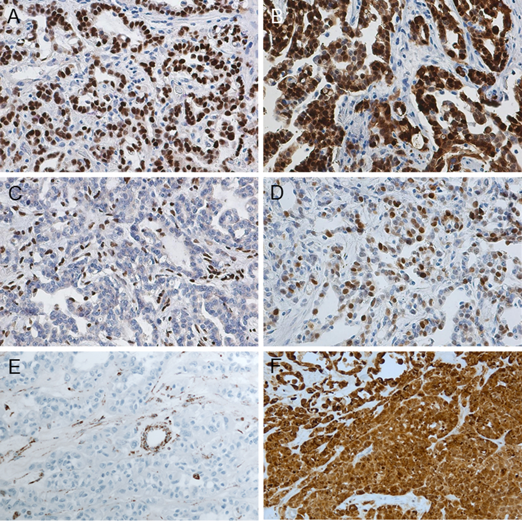
Immunohistochemistry (A-F). (A) PAX8: diffuse nuclear positivity. (B) S100A1: diffuse nuclear and cytoplasmic positivity. (C) SMARCB1/INI1: complete loss in tumor cells (internal control shows positive staining). (D) OCT3/4: nuclear positivity. (A-D: RMC) (E) FH: complete loss in tumor cells (internal control shows positive staining). (F) 2SC: strong and diffuse nucleocytoplasmic positivity (E-F: FH-deficient RCC).