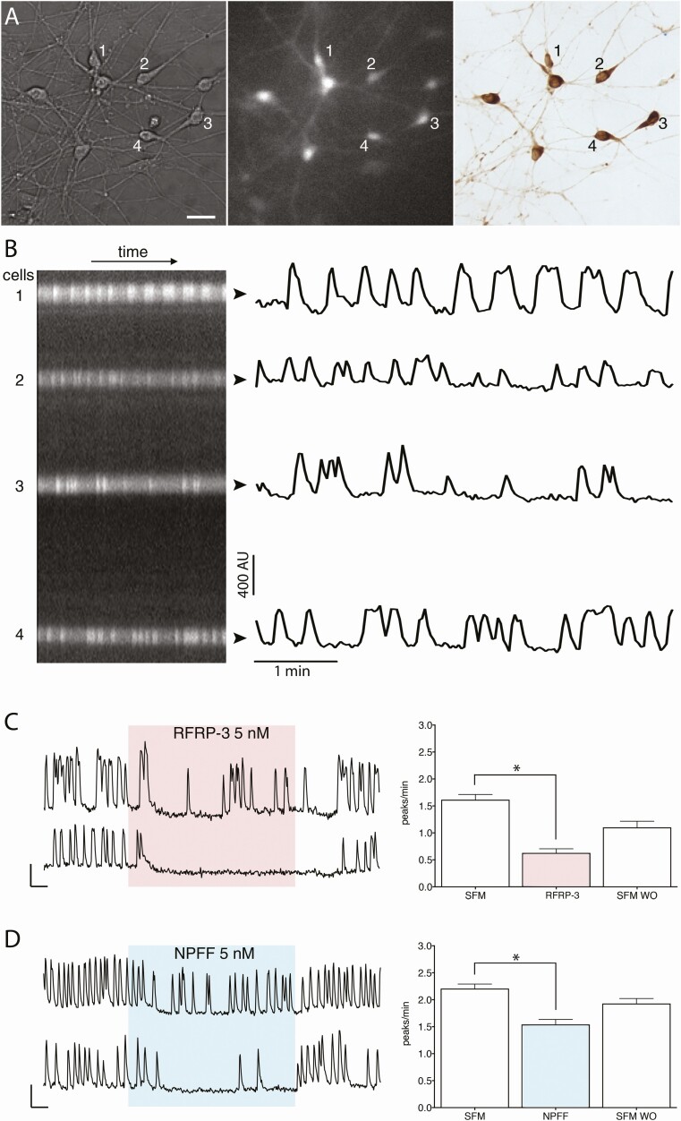
Figure 2.
Imaging of intracellular calcium oscillations in GnRH neurons in explants. (A) GnRH neurons maintained in explants were identified by their fusiform shape (left panel), loaded with calcium green 1 AM (middle panel) and phenotypically confirmed immunochemically post hoc (right panel)(bar 25 µm). (B) Left, kymograph from the cells numbered in A, and, right, corresponding fluctuating optical densities evoked by calcium oscillations. (C,D) Left, 2 representative traces showing calcium oscillations in GnRH neurons after application of RFRP-3 (5 nM) and NPFF (5 nM). Right, Quantification from independent experiments. Both RFRP-3 and NPFF evoked a decrease in the frequency of calcium oscillations. Asterisks indicate P < .05 (paired t-test).

