Figure 6.
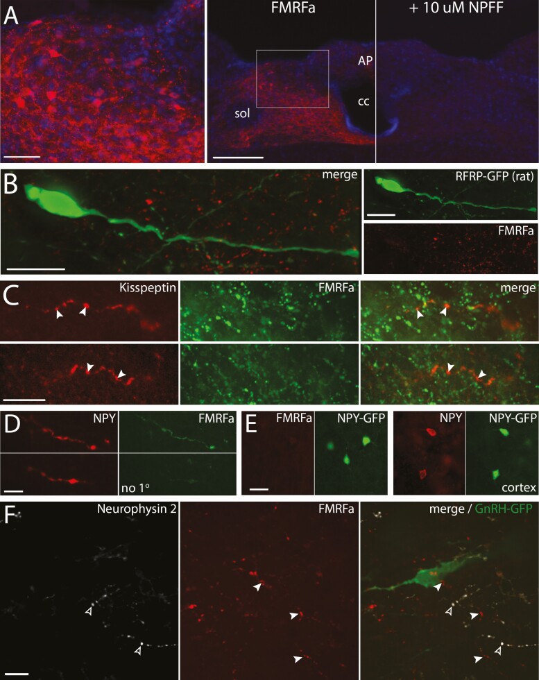
Controls for FMRFa antibody specificity. (A) The FMRFa antisera highlights cell bodies in the dorsal part of the caudal NTS (AP, area postrema; cc, central canal; sol, solitary tract). Left, high magnification confocal image of the boxed area on the middle panel showing cell bodies immunoreacted with FMRFa antisera and counterstained with DAPI (bar 50 µm). Middle, low magnification image containing the left panel, obtained with conventional light microscope (bar 200 µm). Right, low magnification image corresponding to the section in the middle panel, in which the FMRFa antisera was preabsorbed with 10 µM NPFF. (B) Confocal imaging showing the NPFF-like immunoreactivity (FMRFa, red) does not stain RFRP-GFP neurons (green)(rat). Left, merged color channels. Right, individual color channels (bars 20 µm). (C) Confocal imaging showing that the NPFF-like immunoreactivity (FMRFa, green) does not stain kisspeptinergic fibers emerging from the side of the arcuate nucleus (green) (bars 20 µm). (D) Confocal images showing coimmunoreactivity between NPY (red) and NPFF-like (FMRFa, green) in the brainstem. The omission of FMRFa antibody gives no staining in NPY stained fibers with TSA method (bar 10 µm). (E) Confocal images showing no cross-reactivity of the FMRFa antibody with NPY. Left, absence of immunoreactivity with FMRFa in GFP positive neurons in the cortex. Right, presence of immunoreactivity with NPY in GFP positive neurons in the cortex. (bar 20 µm) (F) Flattened stack of confocal images showing no immunoreactivity for neurophysin 2, protein carrier for AVP (white, open arrowheads) in NPFF-like immunoreactive fibers (red, filled arrowheads) surrounding GnRH neuron (green). Left and Middle, individual color channels for neurophysin and FMRFa. Right, merge with GnRH staining.

