Fig. 3.

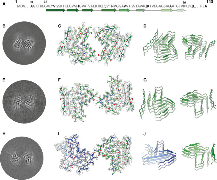
Cryo‐EM structures of type 1 and type 2 filaments with protofilament fold B assembled using seeds from MSA case 1. (A) Primary sequence of α‐synuclein with β‐strands and loop regions shown from dark green (N‐terminal) to light green (C‐terminal). (B) Central slice of the 3D map for type 1 filaments with protofilament fold B. (C) Cryo‐EM density (transparent grey) and fitted atomic model (with the same colour scheme as in A) for type 1 filaments. (D) Cartoon view of three successive rungs of the type 1 filament. (E–G) As (B–D), but for type 2 filaments. (H–I) As (B–D), but for the putative type 2 filament that contains a mixture of protofilament folds A and B.
