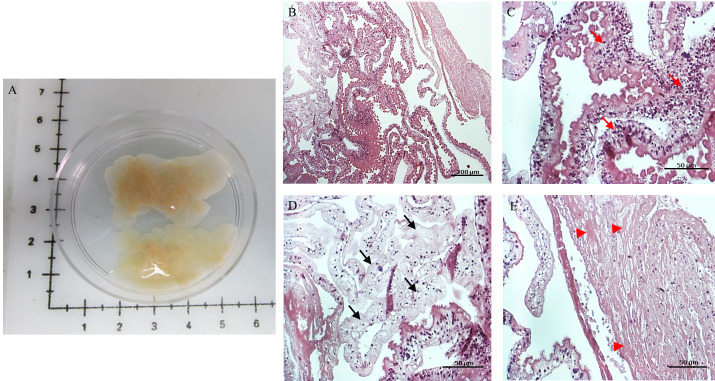
Fig 1. Racemose larvae sample.
Macroscopic view of representative sample (A) and HE stain of the sample (B). Three selected regions with different histological characteristics; region with intact or viable tissue (C), the tegument has normal morphology with microtriches on the surface (red arrows are pointing to hypertrophic/integrity regions); region with tissue degeneration (black arrows) (D); region with a high degree of necrosis (E) characterized by the absence of nuclei (arrow head).

