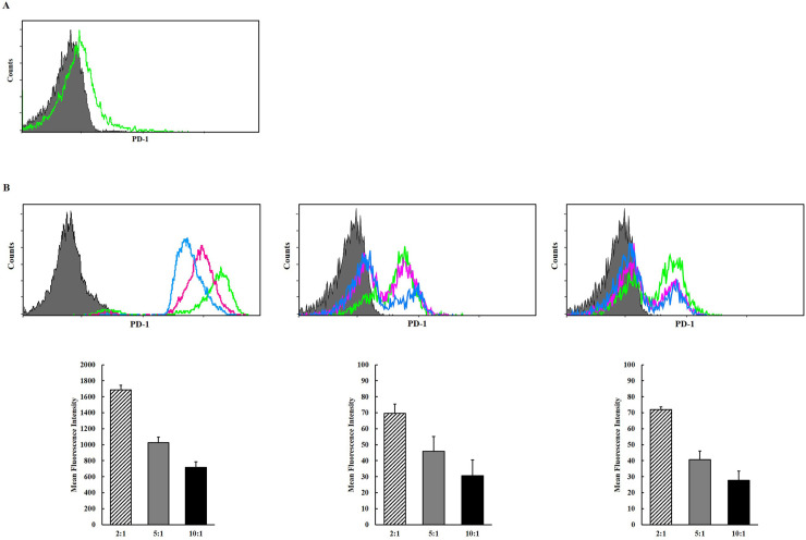
Fig 3.
(A) Surface expression levels of programmed death 1 (PD-1) in steady state NK cells. (B) Surface expression of PD-1 in activated NK-92 cells following co-culture with K562 cells and melanoma cells (A375P, SK-MEL-28), respectively, at each E:T ratio. The upper panel shows representative histograms (green line– 2:1; purple line– 5:1; blue line– 10:1) and the lower panel shows MFIs (diagonal line– 2:1; filled gray– 5:1; filled black– 10:1). The experiments were performed three times.

