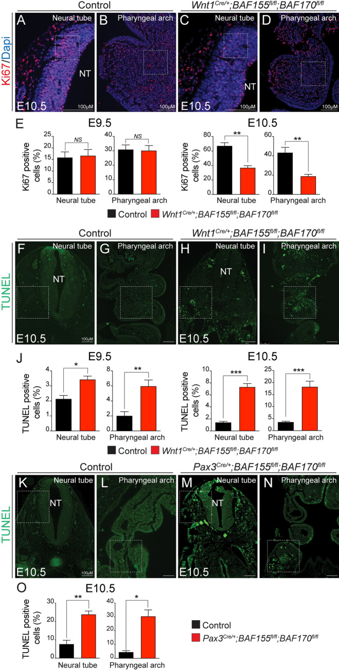
Fig 5. Increased apoptosis and decreased cell proliferation in BAF155/170Wnt1-CKO embryos.
(A-D) Anti-Ki67 immunostaining and Dapi counterstaining on frontal sections with neural tube and pharyngeal arch area of E9.5 and E10.5 control and BAF155/170-deficient (Wnt1Cre/+;BAF155fl/fl;BAF170fl/fl) embryos. (E) Quantification of cell proliferation was calculated as the ratio of Ki67-positive cells to the total number of cells as determined by Dapi counterstaining in the defined area of the neural tube and pharyngeal arch (E). (n = 3–4 each genotype). (F-J) TUNEL assay and quantification was performed on E9.5 and E10.5 control and BAF155/170-deficient (Wnt1Cre/+;BAF155fl/fl;BAF170fl/fl) sections. (n = 3–4 each genotype). (K-O) TUNEL assay and quantification was performed on E10.5 control and BAF155/170-deficient (Pax3Cre/+;BAF155fl/fl;BAF170fl/fl) sections. (n = 3–4 each genotype). Values are reported as means ± SEM (*P < 0.05, **P < 0.01, ***P < 0.001; NS, not significant).

