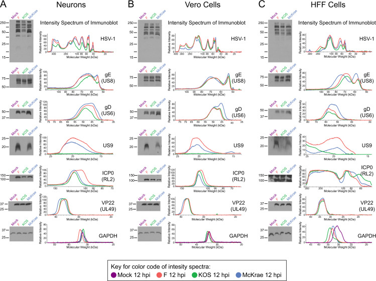
Fig 6. Different viral protein levels between strains demonstrates cell type specificity.
(A) In neurons, immunoblot analysis of total HSV-1 protein, gE, gD, and US9 demonstrates different viral protein levels between strains at 12 hpi. HSV-1 strain KOS exhibited overall lower levels of total viral protein as well as less gD (see also S5 Fig). Additionally, marginally lower gE levels and a complete lack of US9 expression were observed in KOS-infected neurons. In contrast, VP22 protein levels were relatively consistent expression between strains. Subtle differences in total protein, ICP0, gE, gD, and US9 levels between strains were particularly evident in the corresponding intensity spectra. In contrast to the neuronal infections depicted in (A), Vero epithelial cell lysates did not reveal lower levels of total viral protein, gE, or gD in KOS-infected cells (B). Due to the known point mutation in US9 [44–46], this protein is not detected in Vero cells either. Consistent with neurons, no differences in VP22 levels between strains were evident in Vero cells and subtle differences such as higher levels of US9 and ICP0 in strain F persisted in both cell types. (C) Western blots in primary human fibroblasts (HFFs) demonstrate similar trends in viral protein levels as observed in neurons, although to a marginally lesser degree. In HFFs the immunoreactivity of a high molecular weight band at roughly 150 kDa was detected in all lanes, including mock-infected cells, and likely indicates non-specific binding of anti-ICP0 antibody to an HFF-specific cellular protein. Blots shown are representative of 3–4 biological replicates, each prepared and immunoblotted independently. All blots depicted in A–C come from a single one of these biological replicates.

