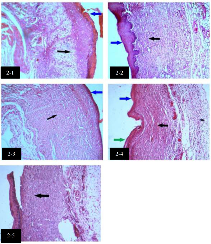
Fig. 2.
Histological differences among groups with H&E staining, on day 7
2-1: Filling of the wound with granulation tissue and irregular collagen fibers (black arrow), no epidermis and presence of superficial clot (blue arrow) in control group on day 7 (H&E, X10).
2-2: Filling of the wound with granulation tissue and regular collagen fibers (black arrow), epidermis formation (blue arrow) in Eucerin-treated group on day 7 (H&E, X10).
2-3: Filling of the wound with granulation tissue and regular collagen fibers (black arrow), epidermis formation (blue arrow) in white tea 5% ointment (Eucerin)-treated group on day 7 (H&E, X10).
2-4: Filling of the wound with granulation tissue and regular collagen fibers (black arrow), Keratinized (green arrow) superficial epidermis formation (blue arrow) in gel-treated group on day 7 (H&E, X10).
2-5: Filling of the wound with granulation tissue and dense regular collagen fibers (black arrow) in white tea 5% gel-treated group on day 7 (H&E, X10).

