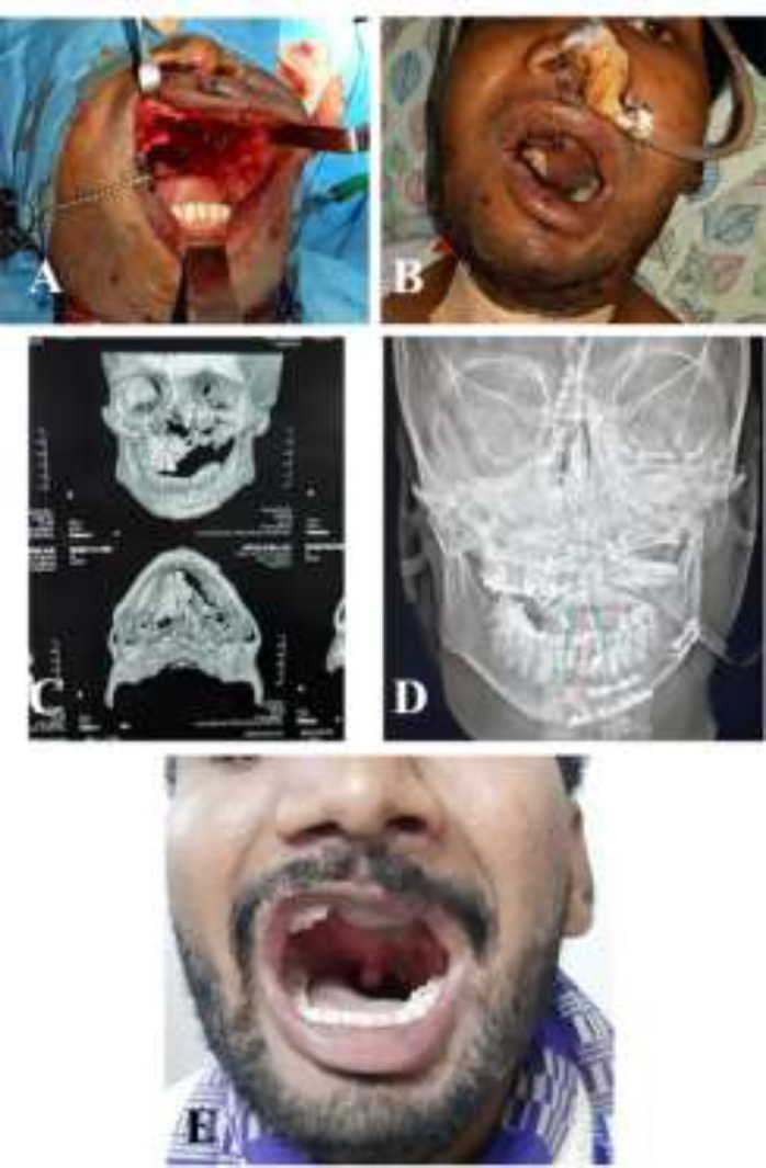
Fig. 4.

A: Post traumatic defect in the maxilla with loss of anterior wall in maxilla, alveolus, and more than half of the palate. B: Early post-operative picture of free fibula osteomusculocutaneous flap with flexor hallucis longus used to fill the antrum. C: Pre-operative CT scan image showing the defect. D: Post-operative X ray film showing the fibula. E: Late post-operative photo showing flap well settled
