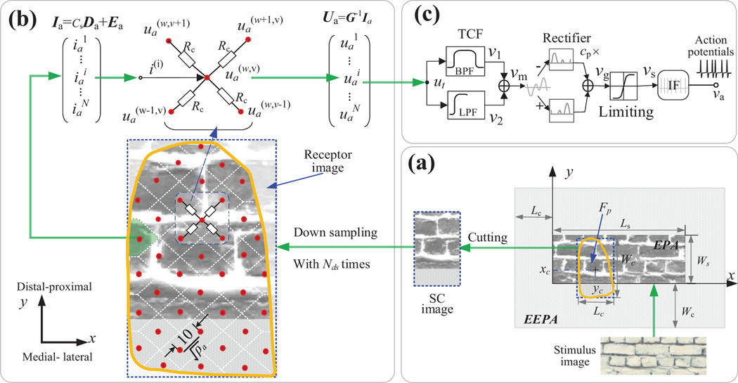
Fig. 1.
Schematic of the whole model. (a) Simplified skin contact (SC) model based on image. (b) The resistance network model of connecting all sampled tactile afferents in fingertip skin area shown as yellow curve box. In the skin area of fingertip, each red dot represents an afferent (node) and was connected by 4 resistors. The white dotted lines in skin area represent the boundaries for dividing the receptor image into several a small area for each tactile afferent in skin area. (c) Model of the single-unit.

