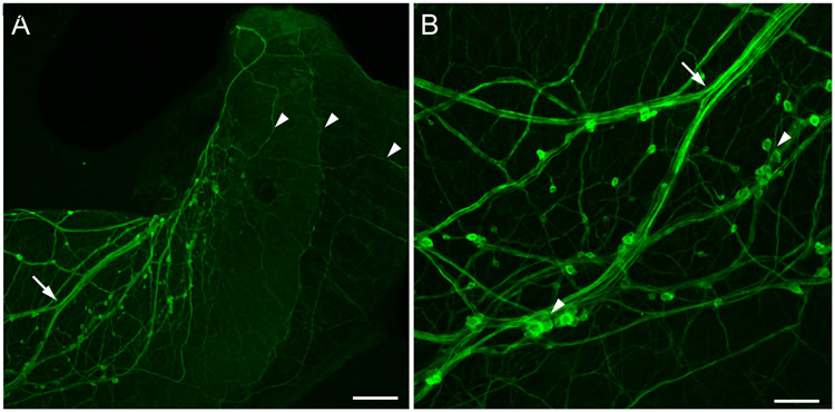
Figure 1.
TH-like immunoreactivity in the albumen gland of the opisthobranch Bursatella leachii. (A) Low power image showing bundles of THli fibers (arrow) entering the albumen gland. Small immunoreactive somata were observed adhering to the fiber tracts. Fine axons (arrowheads) covered the entire surface of the gland. Calibration bar = 200 μm. (B) At higher magnification, some of the small (5 – 10 μm diameter) THli cell bodies were solitary, and others were clustered into small groups (arrowheads). Branch point of the fiber bundle (arrow) corresponds to arrow in panel A. Calibration bar = 50 μm. (Unpublished data; G. Rosado-Mattei and M.W. Miller, Institute of Neurobiology, University of Puerto Rico).

