FIGURE 7.

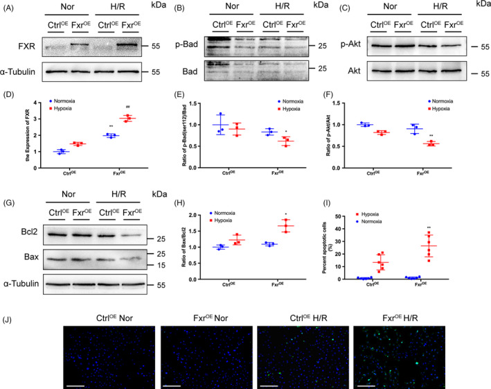
Overexpression of FXR in HK‐2 cells leads to increased cell apoptosis. HK‐2 cells were transfected with FXR plasmid (FXROE) or empty vector (CtrlOE). Cells were treated with wortmannin (Wort) or not and then exposed to hypoxia (1% O2) for 6 h followed by 3 h of reoxygenation. (A) Representative WB images and (D) expression of FXR (**P < 0.01 vs CtrlOE under normoxia, ## P < 0.01 vs CtrlOE after H/R). (B) Representative WB images and (E) expression ratio of p‐Bad to t‐Bad. (C) Representative WB images and (F) expression ratio of p‐Akt to t‐Akt. (G) Representative WB images and (H) expression ratio of Bax to Bcl‐2. (I) Representative images of TUNEL assays (original magnification, ×200; scale bar, 200 μm). (J) Quantitative analysis of apoptosis cells (n = 6 per group). Each column represents the mean ± SD. *P < 0.05 vs siCtrl after H/R, **P < 0.01 vs CtrlOE after H/R
