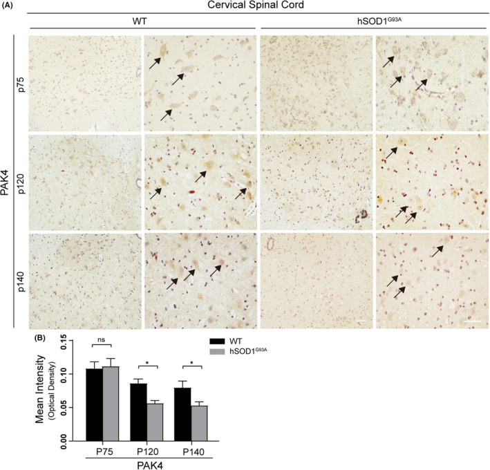
FIGURE 2.

The expression of PAK4 decreased in the cervical spinal cords of hSOD1G93A mice during disease progression. Paraffin sections of cervical spinal cords from hSOD1G93A mice and WT at three stages of the disease were subjected to immunohistochemistry. A, PAK4‐stained MN (maximum diameter ≥ 20 μm) were detected in the anterior horn of spinal cords from both hSOD1G93A mice and WT. Scale bar = 50 μm. B, At stages p120 and p140, OD values of PAK4 significantly decreased in hSOD1G93A mice, while there was no deregulation at stage p75 (n = 3/group, six sections/mouse). Data were provided as means ± SD and were tested for significance using Student's t‐test. ns ≥ .05, *P < .05
