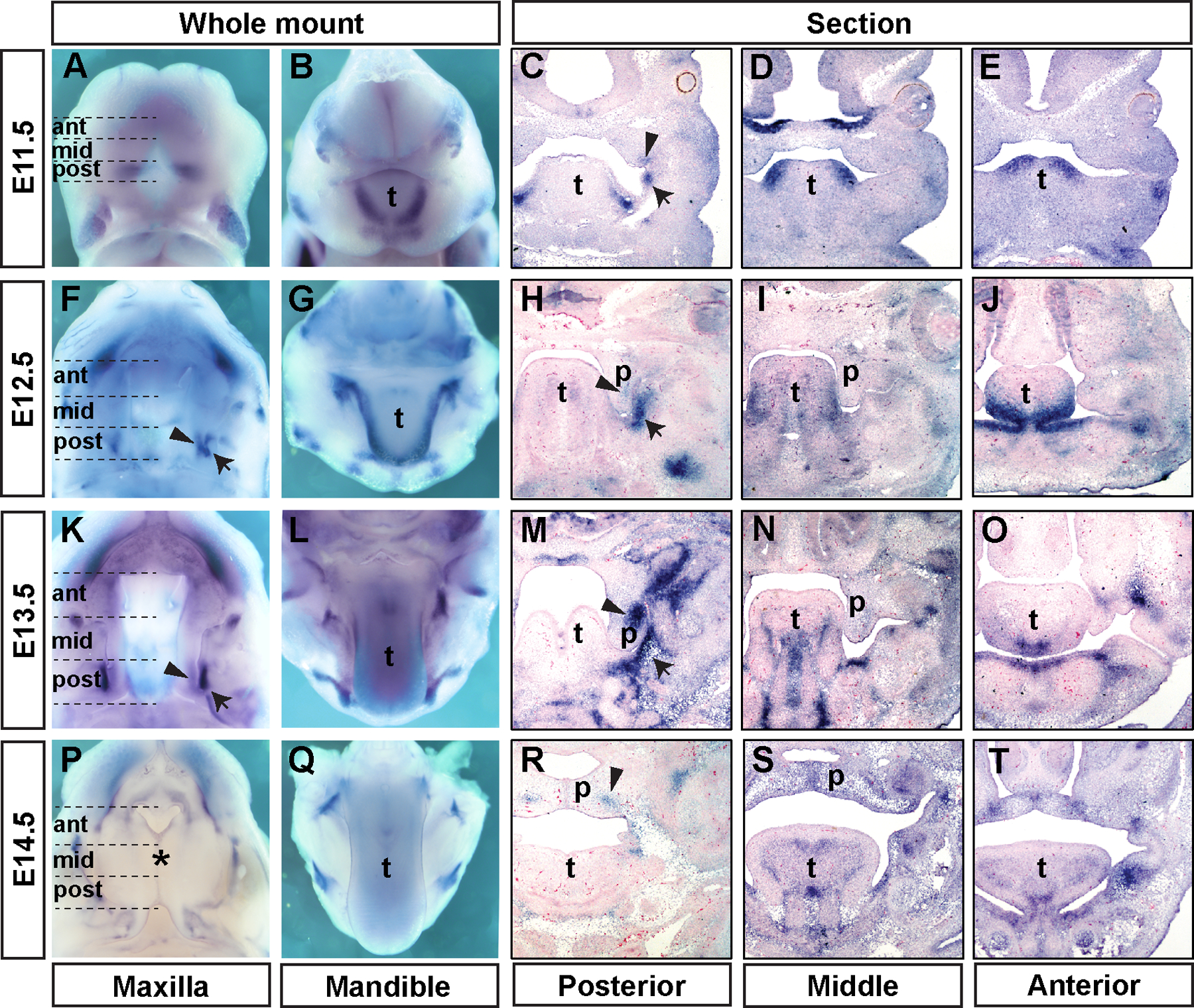
Figure 1. Patterns of Fgf18 mRNA expression during palate development.

Whole-mount oral views of the upper and lower jaws (A-B, F-G, K-L, and P-Q) and frontal sections of mouse embryonic heads (C-E, H-J, M-O, and R-T) showing patterns of Fgf18 mRNA expression in developing mouse craniofacial tissues at E11.5 (A-E), E12.5 (F-J), E13.5 (K-O), and E14.5 (P-T). Representative images of frontal sections from the posterior, middle, and anterior regions of the palatal shelves from each embryonic stage (the regions are indicated on whole mount images in the left column) are displayed from left to right in each row. Arrowheads point to the domain of Fgf18 mRNA expression in the posterior palatal shelf. Black arrows point to the Fgf18 expression at the maxillomandibular junction region, whereas green arrows point to the midline epithelial seam of the fusing secondary palate at E14.5. p, palatal shelf; t, tongue.
