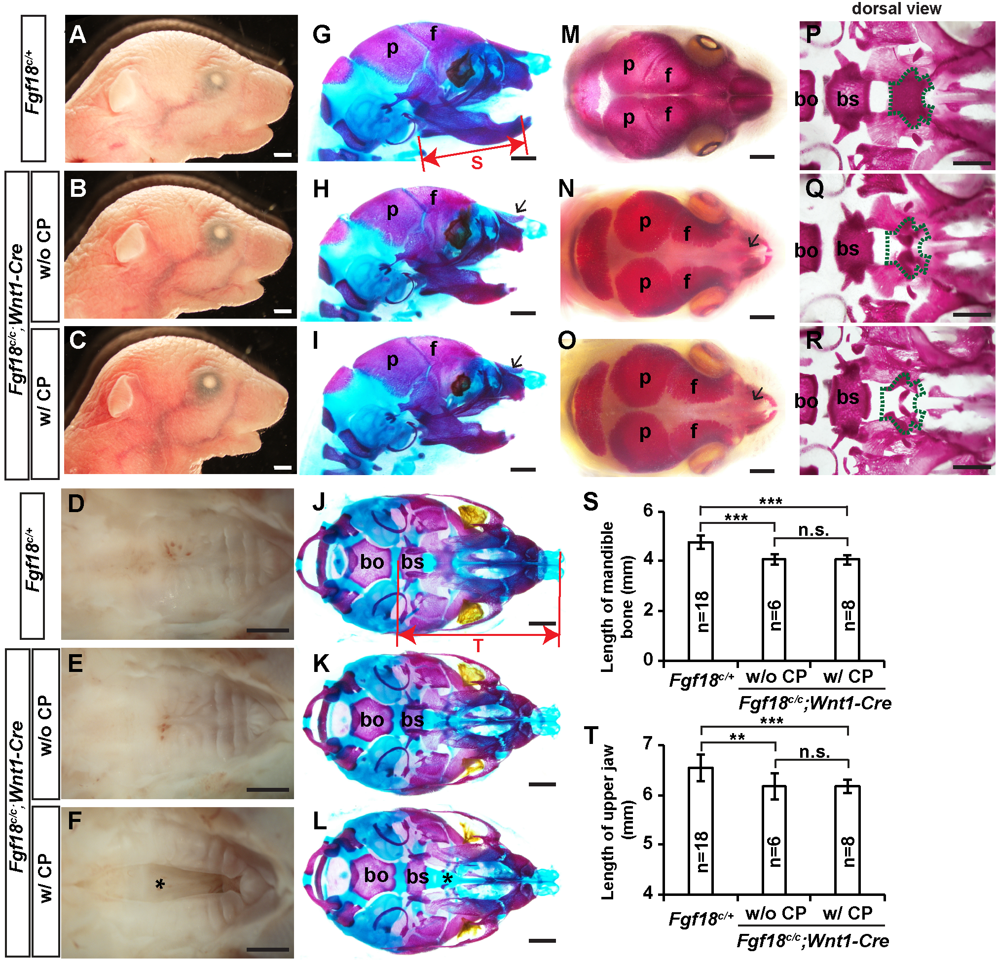
Figure 2. Fgf18c/c;Wnt1-Cre mouse embryos exhibit micrognathia and cranial skeletal defects.

(A-F) Lateral (A-C) and palatal (D-F) views of P0 Fgf18c/+ (A, D) and Fgf18c/c;Wnt1-Cre (B, C, E, F) heads. (G-L) Skeletal preparations of E18.5 Fgf18c/+ (G, J) and Fgf18c/c;Wnt1-Cre (H, I, K, L) embryos were stained with Alizarin red and Alcian blue, with the lateral (G-I) and palatal (J-L) views shown. (M-R) Skeletal preparation of P0 Fgf18c/+(M, P) and Fgf18c/c;Wnt1-Cre (N, O, Q, R) pups were stained with Alizarin red only, with the dorsal views of heads (M-O) and the cranial base (following removal of the calvarial bones) (P-R) are shown. Arrows in H and L point to the underdeveloped nasal bones in Fgf18c/c;Wnt1-Cre mutants. Green dashed lines in P-R show the position of the presphenoid bone. Asterisks in F and L mark the cleft palate. bo, basioccipital bone; bs, basisphenoid bone; f, frontal bone; p, parietal bone. (S and T) Quantitative comparison of the length of the mandibular bone (G) and of the upper jaw (H). **, P<0.01; ***, P<0.001; n.s., no significant difference. Scale bar: 1 mm.
