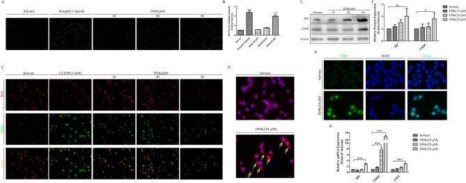
Fig. 4.
HNK can stimulate the mitochondrial damage and endoplasmic reticulum stress. The NB4 cells were incubated with 0, 10, 20, and 30 μM HNK for 24 h. a A fluorescent probe DCFH-DA detected the expression level of ROS in NB4 cells. 0.1 mg/mL Rosup was used as a positive control. b Measured the relative fluorescence intensity of each group of samples using a microplate reader. c Mitochondrial membrane potential was analyzed by JC-1. 0.1 mM CCCP was used as a positive control. d A specific endoplasmic reticulum fluorescent probe (red) was used to observe the formation of endoplasmic reticulum vacuoles (yellow allows) after 30 μM HNK treatment. e, f The endoplasmic reticulum stress protein BiP and CHOP were analyzed by western blotting. g The fluorescence intensity of endoplasmic reticulum stress protein ATF4 (green) was observed by immunofluorescence. h Real-Time PCR was used to measure the mRNA expression of BiP, CHOP, and ATF4. The results are expressed as mean ± SD (n = 3). Compared with the solvent group, *P < 0.05, **P < 0.01, and ***P < 0.001 (Color figure online)

