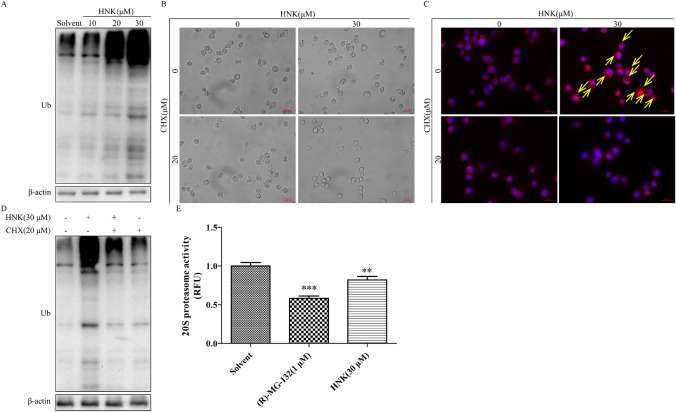
Fig. 5.
HNK increases protein ubiquitination by inhibiting proteasome activity. a The expression of ubiquitinated proteins after HNK treatment for 24 h. NB4 cells were pretreated with 20 μM CHX for 2 h and then incubated with HNK (0 or 30 μM) for 24 h. b Observed cytoplasmic vacuoles with a microscope. c Endoplasmic reticulum vacuoles (yellow allows) were marked using a specific endoplasmic reticulum fluorescent probe (red). d Western Blot analysis was used to measure the level of protein ubiquitination. e NB4 cells were treated with HNK (0 or 30 μM) for 24 h, and the cells were collected for 20S protease activity determination. 1 μM (R)-MG-132 was used as a positive control. The results are expressed as mean ± SD (n = 3). Compared with the solvent group, **P < 0.01 and ***P < 0.001 (Color figure online)

